

Health
Consultant Pathologist takes us behind the microscope for Breast Cancer Awareness Month 2023
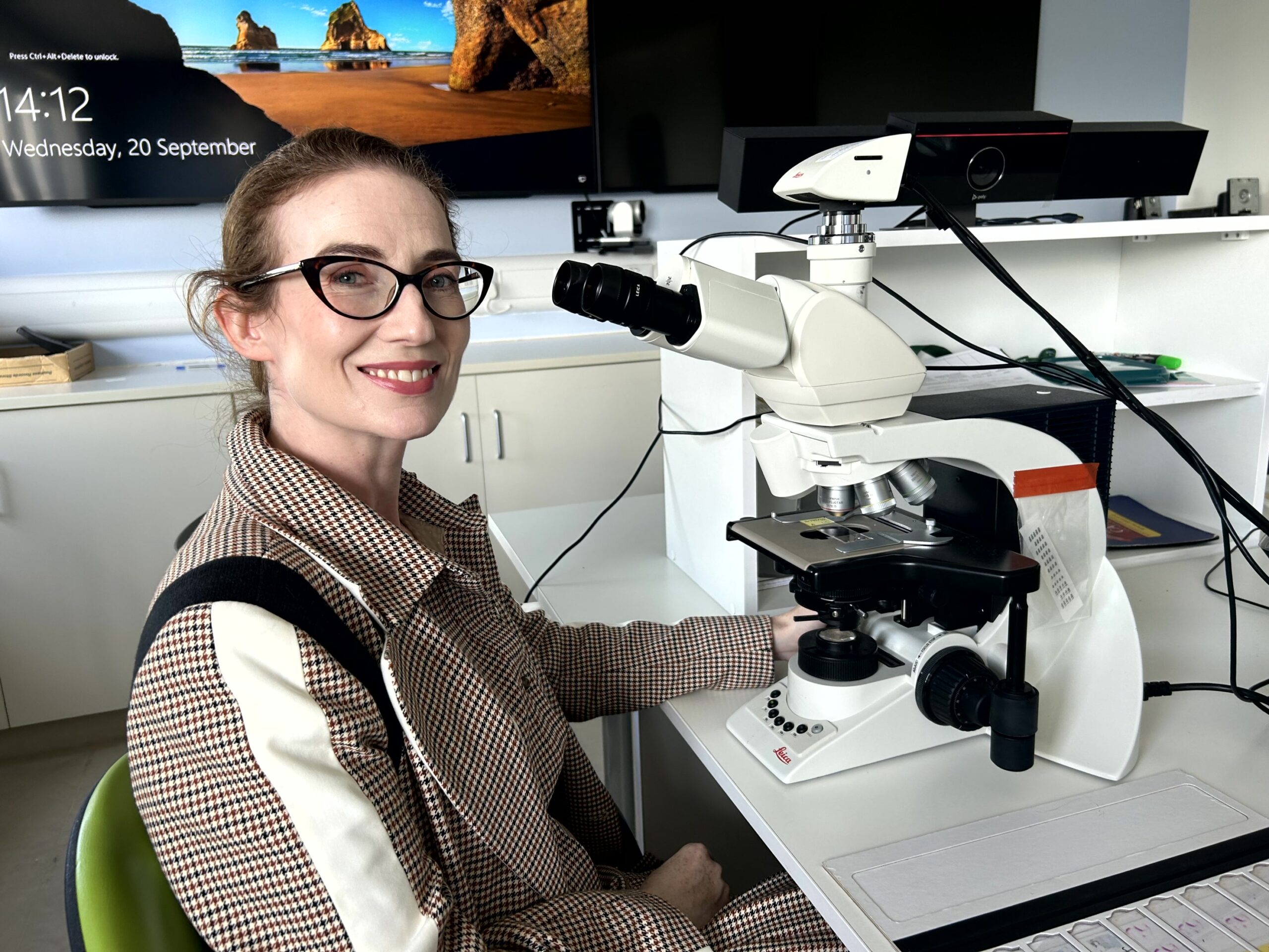
For Breast Cancer Awareness Month this October 2023, this editorial piece by Dr Máire Lavelle, Consultant Pathologist, University Hospital Limerick, answers the question of “what do you actually DO?”
Marking 2023 Breast Cancer Awareness month this October, Dr Máire Lavelle, Consultant Pathologist, University Hospital Limerick, explains her role in the diagnosis of disease and treatment options for women attending the region’s Symptomatic Breast Unit
I am the Lead Breast Consultant Pathologist in UHL. When I tell people that I’m a pathologist, it is sometimes assumed that I attend crime scenes and solve murders. When I explain that I’m not a forensic pathologist, I’m a hospital pathologist, I am often asked: “but what do you actually DO?”
Pathology is the study of disease. As a breast pathologist, I diagnose breast disease under the microscope.
When a patient comes to the breast unit with a problem, they see a surgeon who takes a clinical history and examines them. If necessary, the surgeon may request a scan. If the radiologist performing the scan sees something abnormal, they will take a biopsy of it, which a tissue sample of the abnormal area of the breast which is put in a container of fixative and sent to the Histopathology laboratory.
advertisement

advertisement

advertisement

We can’t just put the biopsy straight under the microscope. The microscope light must pass through the sample to be seen, so the sample must be cut very thinly so light can pass through it and it can be assessed.
To achieve this, the tissue sample is fixed in formalin in its container. It is then embedded (either by a pathologist or a medical scientist) in a small cassette and covered in paraffin wax, creating a wax block.
The tissue fixed in the wax block is cut into extremely thin strips by a medical scientist on a machine called a microtome. The thin strips are placed onto a glass slide.
At this point, the cells are still difficult to see, because they are very transparent, so the slide goes through a staining machine where tissue dyes are applied to the sample which colour the cells so they can be seen. The dyes used are haematoxylin and eosin, which dye the tissue in pinks, blues, reds and purples.
The glass slide is dried and then a glass coverslip is glued onto the top of the sample, so the sample is trapped firmly between two slides of glass.
This is the point when I can put the slide under the microscope and make an assessment.
Sometimes I can make an immediate diagnosis. Sometimes the interpretation is more complex and I might need to perform more chemical tests on the sample to establish a diagnosis.
Even if the sample is clearly cancerous, I may still need to perform more tests to establish the subtype of cancer and what hormones it expresses. All these extra tests allow decisions to be made on potential future surgery, chemotherapy and radiation therapy.
The pathological diagnosis under the microscope is not the end of my duties for this patient. Each breast sample is discussed at the breast multidisciplinary meeting. Surgeons, radiologists, pathologists, oncologists, radiation oncologists and breast care nurses all attend this meeting and we each discuss our findings to establish the diagnosis for the patient and discuss the treatment options for each patient.
My job also involves cancer research as well as education and training of medical students, junior doctors and medical scientists. The smooth running of the laboratory requires a lot of work from the entire laboratory team of which I am a part.
What is my favourite part of the job? That is a difficult question! Pathology is often described as “the bridge between science and medicine” and I really enjoy having a hand in both fields. There is huge job satisfaction in being part of the breast team to treat patients with breast cancer. That is why I became a doctor: to help people.


Mountains, Fall on Us by artist Austin McQuinn runs at Limerick City Gallery of Art until June 9, 2024
PHOTOS Vacious by Sinead O’Brien enters ‘The Body Suit Era’ with launch of new outerwear collection

Milford Care Centre and Rugby Legend Keith Earls honoured at Limerick Person of the Year Awards 2023

Karen Fitzgibbon’s ‘The World’s End’ set for O’Mahony’s launch on Thursday, May 2
Bedford Row Family Support Project celebrates 25 years as a beacon of hope to Limerick families

Broadford community comes together to support Carol Liston O’Connor following Motor Neurone Disease diagnosis
PHOTOS Limerick creatives come together for A Special Night for Gaza fundraiser at Dolans, Sunday, April 7

Celebration of verse as April is Poetry Month in Limerick
